Blog, News & Offers
OCT technology, seeing deeper into the eye.
Posted on Saturday, July 27, 2019
At Colin Lee Opticians we have always prided ourselves on providing a first class service and leading the way in innovative technologies within eye health care. We were one of the first optical practices to introduce OCT (Ocular Coherence Tomography) technology to the high street a few years ago and we now expanding this tech to more of our practices. This has allowed for earlier detection of many eye conditions including glaucoma and macular degeneration as well as providing peace of mind to patients to patients on NHS waiting lists.
What is OCT and how does it work?
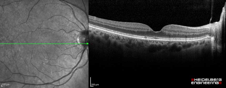
Ocular Coherence Tomography is a non-invasive imaging scan. It is the optical equivalent of ultrasound: ultrasound uses sound waves to bounce of layers of tissue of varying reflectivity to build up an image; OCT works in a similar way. We can then see a cross-sectional image of the retina, thickness measurements are taken and maps formed, which are all important in diagnosing and monitoring eye diseases.
The retina is the light sensitive membrane at the back of the eye that detects images and lets us see – a bit like the film in a camera. With OCT your optometrist can see each of the retina’s distinctive layers enabling earlier detection of many eye conditions.
We all love cake and an iced cake is a great way to think about how OCT works. The top layer of the retina is the icing and without OCT (such as with a microscope or looking into the eye with standard equipment or with retinal photography) we can only see the icing without being able to see inside the cake. Are the layers all even? Is there the right amount of jam, or has it leaked into the sponge? Is there anything hiding in the layers of that cake? Without looking inside the cake, only looking at the icing, you just don’t know. Luckily we don’t need to cut into the cake/eye to find out what is going on in the layers under the surface of the retina as the OCT simply shows us on the screen.
What happens during an OCT appointment?
During an OCT scan you will sit in front of the OCT equipment and rest your head on a support to keep it as still as possible. The camera will then scan your eye, without touching it. You will simply see a few red lines moving around and you will be asked to looks at a green or white dot. Scanning takes about 5 – 10 minutes in total – but most of this time is getting you positioned correctly – the actual scan itself is just a couple of minutes. If you have an OCT scan immediately preceding your eye examination then the optometrist will be able to show you the fascinating images on the screen in the consulting room. Most people don’t require any drops to have the scan either, but the odd few have too small pupils to get a good view so it would be in their
interest to have the drops put in for the quality of the scan.
Why should you have an OCT scan?
OCT scans can identify many more conditions, in addition to glaucoma and macular degeneration (both wet and dry), conditions such as macular holes, macular pucker, macular oedema, central serous retinopathy, diabetic retinopathy & vitreous traction. Many patients choose to have a scan when referral is advised to the Hospital eye clinic, to firstly get answers faster rather than waiting several months for an appointment at the hospital and secondly decrease the For many conditions (and also for routine eye health checking), serial scans give us the most valuable information. If we can build up a number of images over time, it allows us to see if a condition is developing or changing. In addition in detecting disease, serial OCT scans carried out at each eye examination can also give reassurance that healthy eyes are continuing to be perfectly healthy!
We have an OCT at our Aldridge and Tamworth (Jenks) practices so far, so all within easy reach of our other 5 practices.
Blog Search
Blog Categories
- News (62)
- Sunglasses (3)
Blog Archives
- March2024 (1)
- November2023 (1)
- October2023 (1)
- August2023 (2)
- June2023 (2)
- April2023 (1)
- November2022 (1)
- October2022 (2)
- September2022 (1)
- August2022 (4)
- June2022 (3)
- May2022 (3)
- March2022 (3)
- December2021 (1)
- November2021 (2)
- July2021 (2)
- June2021 (1)
- May2021 (2)
- April2021 (3)
- March2021 (2)
- February2021 (4)
- January2021 (1)
- December2020 (1)
- November2020 (2)
- October2020 (1)
- September2020 (2)
- July2020 (1)
- April2020 (2)
- March2020 (3)
- January2020 (1)
- July2019 (3)
- June2019 (2)
- February2019 (1)
- October2016 (1)
- September2016 (1)
- June2016 (1)
Offices
- Aldridge (1)
- All Offices (24)
- None (37)